What Is a Dental Bitewing X-Ray? How Dentists Detect Hidden Problems Between Teeth
Learn what bitewing dental X-rays show, how they help detect cavities between teeth, and why dentists use them during exams in National City, CA.

You brush twice a day, floss regularly, and visit your dentist for routine checkups. Yet, some dental problems can develop without causing noticeable symptoms. Cavities between teeth, for example, may remain hidden because the affected surfaces are difficult to see during a visual examination.
This is one reason dentists sometimes recommend bitewing X-rays. These dental images help reveal areas between the teeth that may not be visible during a regular oral exam. They can also provide information about the bone supporting your teeth.
Understanding what bitewing X-rays show, when they may be needed, and how they fit into preventive dental care can help you make informed decisions about your oral health.
At Crown Dental Group in National City, California, dental imaging may be used as part of an individualized examination when it can provide useful diagnostic information.
What Is a Bitewing Dental X-Ray?
A bitewing X-ray is a type of dental radiograph that captures images of the crowns of the upper and lower teeth in a particular section of the mouth. The patient typically bites down on a small holder or positioning device to help keep the image receptor in place.
The term bitewing comes from the traditional technique of using a small tab that the patient bites on during image capture.
Unlike some other dental X-rays, which focus on the roots of individual teeth or broader areas of the jaw, bitewing images are primarily used to examine the crowns of the teeth and the spaces between them. They can also show the level of the bone around the teeth in the imaged area.
Dentists commonly use bitewing X-rays to help identify tooth decay between neighboring teeth, assess certain existing restorations, and evaluate bone levels when clinically appropriate.
These images supplement a clinical examination. They do not replace the dentist’s assessment of your teeth, gums, symptoms, and dental history.
What Can a Bitewing X-Ray Detect?
Bitewing X-rays can reveal several findings that may be difficult to identify through a visual examination alone.
1. Cavities Between Teeth
One of the main reasons dentists take bitewing X-rays is to check for interproximal caries, which is tooth decay that develops on the surfaces where neighboring teeth touch or sit close together.
These areas can be difficult to inspect directly, even when you brush and floss consistently. Early decay may not cause pain or produce an obvious visible change.
On a bitewing image, a dentist may identify changes in the tooth structure that suggest decay. The appearance, location, and extent of these changes help the dentist determine whether further assessment or treatment is appropriate.
Not every area that appears suspicious on an X-ray requires the same treatment. The dentist considers the image alongside the clinical findings and the patient’s risk factors before recommending the next step.
2. Decay Beneath or Around Existing Fillings
Bitewing images may help dentists evaluate certain areas around existing restorations, including fillings and crowns, when those areas are visible on the image.
For example, the dentist may look for signs of decay near the edges of a restoration or other changes in the surrounding tooth structure.
However, X-rays cannot identify every problem beneath or around a restoration. Some defects may require direct examination or additional diagnostic methods.
If you have an older filling or crown, your dentist may consider its condition, your symptoms, and your previous dental history when deciding whether imaging is appropriate.
3. Bone Levels Around the Teeth
Bitewing X-rays can show the height of the alveolar bone, which supports the teeth. This information may help dentists evaluate bone levels when assessing periodontal health.
Changes in the supporting bone can be associated with periodontal disease, although an X-ray alone does not establish the complete diagnosis.
Your dentist may also measure gum pockets, assess bleeding and inflammation, review your medical history, and examine the teeth and surrounding tissues.
When considered together, these findings can help determine whether additional periodontal evaluation or treatment is needed.
4. Problems That May Not Cause Symptoms Yet
Dental decay and some other changes can develop before a person notices pain or sensitivity. Bitewing X-rays may help identify certain abnormalities at an earlier stage, depending on their location and extent.
This does not mean that every dental problem will appear on an X-ray or that imaging can predict all future issues. Very early changes may be difficult to detect, and some conditions are better evaluated through a clinical examination or different imaging techniques.
The purpose of bitewing imaging is to provide additional information that helps the dentist make a more informed assessment.
What Does a Bitewing X-Ray Show Compared With Other Dental X-Rays?
Different dental X-rays are designed to answer different diagnostic questions. The type selected depends on the area being evaluated and the information the dentist needs.
| Type of dental X-ray | Common purpose |
|---|---|
| Bitewing X-ray | Examines the crowns of upper and lower teeth in the same area, including spaces between teeth and nearby bone levels |
| Periapical X-ray | Shows an entire tooth, including its root and the surrounding area |
| Panoramic X-ray | Provides a broad view of the teeth, jaws, and surrounding structures |
| Cone beam CT (CBCT) | Produces three-dimensional images for selected diagnostic and treatment-planning needs |
A bitewing image is particularly useful when the dentist needs to investigate possible decay between teeth. A periapical image may be more useful when evaluating a tooth’s root or the surrounding tissues. Panoramic imaging and CBCT serve different purposes and are not automatically necessary for routine dental examinations.
Your dentist should select the imaging method that best addresses the clinical question rather than using every available type.
What Happens During a Bitewing X-Ray?
For most patients, taking bitewing X-rays is a quick and straightforward process.
Step 1: Positioning the Image Receptor
The dental team places a small digital sensor or another image receptor inside the mouth. A positioning device helps align it with the teeth being examined.
You may be asked to close your teeth gently around the holder to keep it steady.
Step 2: Taking the Image
The dental X-ray equipment is positioned outside the mouth. The dental team captures the image while you remain still for a brief moment.
You may need more than one image to examine different sections of the mouth. The number depends on the area being assessed and the quality of the images required.
Step 3: Reviewing the Results
The dentist reviews the images for signs of decay, changes around restorations, bone-level concerns, or other findings relevant to your examination.
If an area looks unusual, the dentist may examine it more closely, compare it with earlier images, or recommend additional assessment.
Does a Bitewing X-Ray Hurt?
Taking a bitewing X-ray should not be painful. You may feel pressure from the sensor or positioning device, particularly if you have a sensitive gag reflex or a small mouth.
Tell the dental team if the holder feels uncomfortable. They may be able to adjust its position or use a different technique to improve your comfort.
Are Bitewing Dental X-Rays Safe?
Dental X-rays use ionizing radiation, so they should be taken when the expected diagnostic benefit justifies the exposure. Modern dental imaging uses relatively low radiation doses, and digital systems can support efficient image capture.
The dental team should follow appropriate radiation-safety practices, including using suitable positioning and exposure settings. The need for imaging should be determined individually rather than based on a fixed schedule for every patient.
If you are pregnant or think you may be pregnant, tell your dentist before imaging. Your dentist can consider the urgency of the diagnostic question, available alternatives, and appropriate safety measures.
For children and adults alike, the goal is to obtain the necessary diagnostic information while avoiding unnecessary exposure.
How Often Do You Need Bitewing X-Rays?
There is no single schedule that is appropriate for every patient. The recommended frequency depends on your likelihood of developing cavities, your dental history, your current oral health, and whether previous images show areas that need monitoring.
Your dentist may recommend bitewing X-rays more frequently if you have a higher risk of tooth decay or a history of cavities between teeth. If you have a low risk of decay and your previous examinations have been stable, imaging may be needed less often.
Other factors can influence the decision, including existing restorations, changes in symptoms, and the time since your last suitable images were taken.
A dentist should review your individual circumstances before recommending new X-rays. If you have recent dental images from another office, let the dental team know. Those images may be useful if they are available, sufficiently clear, and appropriate for your current needs.
Can You Have Cavities Even If Your Bitewing X-Rays Look Normal?
Yes. Bitewing X-rays are useful diagnostic tools, but they have limitations.
Very early decay may not be visible on an image. Problems on surfaces that are poorly captured or obscured may also be difficult to evaluate. In addition, dental conditions such as gum inflammation, soft-tissue changes, and some cracks require other methods of examination.
This is why dentists combine imaging with a clinical assessment. They may examine the teeth directly, check for symptoms, assess the gums, and consider your risk factors.
If you have persistent tooth pain, sensitivity, swelling, or discomfort when chewing, tell your dentist even if previous X-rays were normal. Your symptoms may require a different or more focused evaluation.
How Can You Reduce Your Risk of Cavities Between Teeth?
Bitewing X-rays can help identify certain areas of concern, but daily preventive habits remain important for reducing the risk of tooth decay.
Consider the following practices:
- Brush twice a day: Use fluoride toothpaste and brush all tooth surfaces carefully.
- Clean between your teeth daily: Dental floss or an appropriate interdental cleaner can help remove plaque and food debris from areas a toothbrush may not reach effectively.
- Limit frequent exposure to sugary foods and drinks: Repeated sugar exposure can contribute to the development of tooth decay.
- Drink water regularly: Water helps support normal oral function, and fluoridated tap water can contribute to cavity prevention where available.
- Attend recommended dental visits: Regular examinations allow your dentist to assess your oral health and recommend preventive care based on your needs.
- Discuss your cavity risk: If you frequently develop cavities, ask whether additional preventive measures, such as fluoride treatments, may be appropriate.
Even with good home care, cavities can still develop. Regular assessment helps identify concerns that may need attention.
Schedule a Dental Examination at Crown Dental Group
Bitewing X-rays help dentists evaluate areas between the teeth that may be difficult to see during a visual examination. They can be particularly useful for detecting certain types of tooth decay and assessing the bone around the teeth.
However, these images are only one part of a comprehensive dental assessment. The decision to take them should reflect your oral health, dental history, symptoms, and individual risk factors.
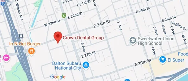
If you need a dental examination in National City, CA, Crown Dental Group can assess your needs and explain whether dental imaging may be beneficial. Patients from Chula Vista, Bonita, Imperial Beach, and surrounding South Bay San Diego communities can contact the office to arrange a visit.
- Crown Dental Group
- 2405 Transportation Ave
- National City, CA 91950
- Phone: (619) 474-6200
- Email: info@crowndentalnc.com
Frequently Asked Questions
1. What is the main purpose of a bitewing X-ray?
A bitewing X-ray primarily helps dentists examine the crowns of upper and lower teeth in the same area. It is commonly used to detect cavities between teeth and assess certain bone levels around the teeth.
2. Can a bitewing X-ray detect cavities between teeth?
Yes. Bitewing images can reveal changes in tooth structure that suggest decay between neighboring teeth. The dentist interprets these findings alongside the clinical examination before deciding whether treatment is necessary.
3. How many bitewing X-rays do I need?
The number depends on the areas that need to be examined, the quality of the images, and your individual diagnostic needs. Your dentist will determine how many images are appropriate.
4. Are bitewing X-rays safe for children?
Dental X-rays may be appropriate for children when they provide useful diagnostic information. The dentist considers the child’s age, cavity risk, dental development, and clinical findings before recommending imaging.
5. Do bitewing X-rays show the roots of teeth?
Bitewing images primarily show the crowns of the teeth and the areas between them. They may show portions of the roots, but they are not designed to display the full length of each root. A periapical X-ray may be more appropriate when the dentist needs to evaluate the entire tooth and surrounding root area.
6. Can I refuse a bitewing X-ray?
You can discuss any dental procedure with your dentist, including X-rays, and ask why it has been recommended. Your dentist can explain the potential benefits, limitations, and alternatives. Together, you can make an informed decision about your care.
7. Do I need bitewing X-rays at every dental cleaning?
Not necessarily. The need for X-rays depends on your dental history, cavity risk, previous images, and current clinical findings. Some patients need them more frequently than others.
8. What if my bitewing X-ray shows a cavity?
Your dentist will evaluate the location and apparent extent of the decay alongside the clinical examination. Depending on the findings, the recommendation may involve monitoring an early lesion, improving preventive care, or restoring the tooth with a filling or another appropriate treatment.
Find us here